Nanoscale secondary ion mass spectrometry (NanoSIMS) is a highly sensitive chemical analysis technique for acquiring 2D and 3D maps of elemental distribution information with exceptionally high spatial and mass resolutions.
It is typically used to produce images of elemental concentration.
NanoSIMS is a powerful analytical technique designed for ultra-high-resolution elemental and isotopic imaging at the nanometer scale. The process begins with a fine-focused primary ion beam—typically less than 50 nanometers in diameter—composed of ions such as cesium (Cs⁺) or oxygen (O⁻). This beam is rastered, or systematically scanned, across the surface of a sample. As the primary ions strike each pixel of the sample, they sputter secondary ions from the surface. These secondary ions are then collected and analyzed, allowing NanoSIMS to construct detailed, high-resolution maps of elemental and isotopic distributions.
A key feature of NanoSIMS is its exceptional spatial resolution, capable of imaging features as small as 50 nanometers. This is achieved through a coaxial lens system that positions the ion probe extremely close to the sample surface. The primary ion beam strikes the sample at a perpendicular angle (90°), while the secondary ions are extracted back through the same optical path, minimizing distortions caused by surface topography or shadowing.
Our Process
Sample preparation
Samples must be flat, dry, and conductive. Metals are polished; biologicals are fixed, embedded, and coated.
Ion sputtering
A focused Cs⁺ or O⁻ beam scans the surface. It ejects secondary ions from each point.
Mass detection
Secondary ions are collected and sorted by mass. Up to 7 ion types are detected at once.
Imaging
Ion data is used to create high-res maps. These show element and isotope locations down to 50 nm.
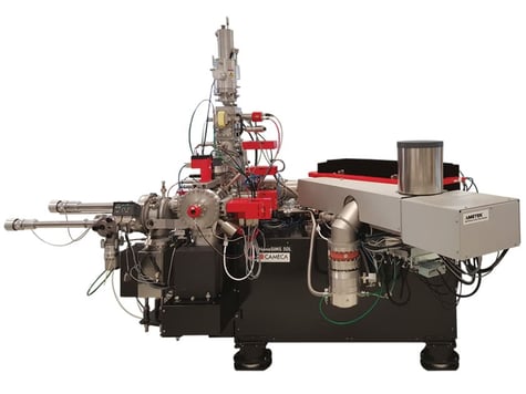
NanoSIMS Instrument Used
Sensitivity: ppm to 1 at.% depending on element
Depth Range: from several 10’s of nm to ~100 microns
Depth Resolution: 10-15 nm
Elements Detected: H – U
Primary Ion: O- or Cs+
Beam Diameter: 50 nm
Mass Analyzer: Magnetic Sector
Number of detected ions in multicollection: 7, analyzed simultaneously
Strengths
1D (line-scan), 2D and 3D element maps can be collected
High mass resolution for isotope analysis
Elemental sensitivity much better than EDS or XPS for trace element analysis
Limitations
Relatively low depth resolution (10-15 nm) compared to other SIMS methods due to the higher primary ion accelerating energy in NanoSIMS
Quantitative analysis requires reference sample(s) to be prepared
Your Material Analysis Begins Here
Our team of experts is ready to provide a detailed and accurate quote tailored to your specific project requirements